| Model: | PM-V6T |
|---|---|
| Brand: | PlenMED |
| Origin: | Made In China |
| Category: | Electronics & Electricity / Other Electrical & Electronic |
| Label: | ultrasound scanner , veterinary equipment , diagnostic apparatus |
| Price: |
US $16059.84
/ pc
|
| Min. Order: | 1 pc |
Product Description
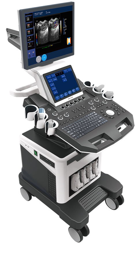
T6-VET
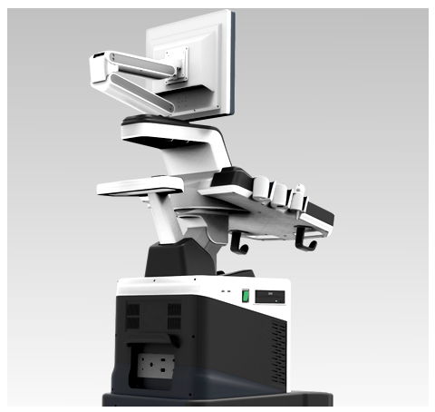
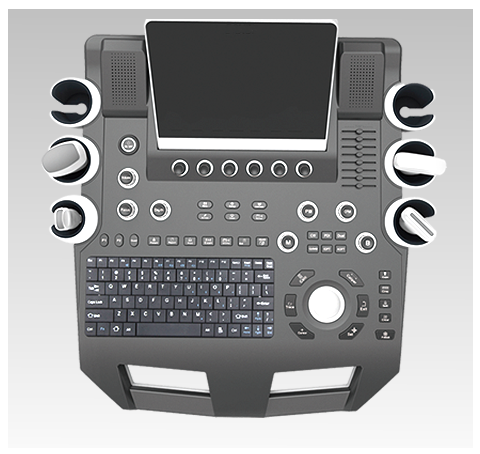
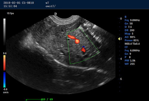
Dual screen ultrasound machine / trolley color doppler ultrasound machine / real time 4D ultrasound
Main Features:
Mode of display
1. Black-and-white image:
B, 2B, 4B, right/left B|M, B|D, right/left PW(D), top/down PW(D), M, B mode local amplification, linear probe trapezoidal imaging, 3D reconstruction/real-time 3D imaging
2. Blood flow images:
Spectrum: B/BC, BC/B, B/D, B/C/D CFM: B|C|D, B|C|M, B|C double real-time, PW(D)(D), CFM, CPA, CW
3. Color Doppler:
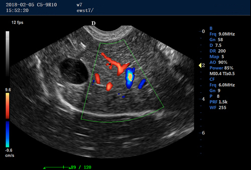
Mode of display: energy, velocity, variance (in support of velocity variance), frequency, gain
Display control: the size and position of the sampling box are both adjustable, the emitting angle of the linear probe is adjustable.
4. Spectral Doppler:
- Sampling volume and position are adjustable
PW(D) tracing supports maximum and average velocity. The threshold of the maximum velocity is adjustable. - Scanning scope: above base-line, below base-line and entire scanning.
PW(D) sound can be turned on or off, and can be regulated in volume.
Velocity measuring: in support of maximum and minimum velocity - Parameters for control of blood flow color images: Doppler frequency, position and size of sampling box, base line, color gain, deflection angle, wall filter, accumulative number of times, etc.
Focusing:
- Electronic focusing + sound lens focusing
- Emission involves single focus, double focuses, triple focuses, and quadruple focuses Continuously dynamic focusing in receiving
Signal processing/Doppler sound
- Dynamic wave filtering
- Quadrature demodulation
- Total gain control
- Composite gain technology: 8-section TGC and D-AGC.
- Low-pass filter
- Secondary sampling
- Acoustic output adjustment
- Adjustable Doppler stereophonic acoustic output
Image processing
- Dynamic scope variation and logarithmic compaction
- Time filtering
- Space filtering
- Frame average
- Fringe enhancement
- Gray scale variation
- B|M- or M-mode scanning speed
- Linear density control
- Scanning angle/width control
- Image optimization
- Upward and downward Image flipping
- Multi-angle image rotation
- Options for color gray-scale bar inversion
- Combined processing of tissue and flow images
- Resolution of black-and-white images: 1024×768×8bits (256 gray-scale)
- Resolution of color images: 8bit × 8bit × 8bit color coding
Obstetric measurement
- With 4 varieties of obstetric databases for reckoning gestation age
- 3 non-editable versions: Asian, European and American, plus 1 editable version: user-customized.
- The gestation age can be reckoned by the information in each version on gestational sac diameter, crown-rump length, biparietal diameter, head circumference, abdominal circumference, femur length, humerus length, abdominal transversal diameter, length of vertebrae, occipital-frontal diameter, cerebellum diameter, fibula length, radius length, Cisterna Magna, inner orbital diameter, output orbital diameter, amniotic fluid index, fetal heart rate, nuchal transparency, distance and area, etc.
- Obstetric report
- Measurement and calculation of amniotic fluid index (AFI)
- Ratio calculation (BPD/OFD, FL/AC, FL/BPD and HC/AC)
- Fetus weight estimation
- Reckoning the gestation age and EDD by LMP and BBT
- Fetal biophysical score
- Fetal growth curve
Multi-fetus detection and measurement: Detection of multiple fetuses
Small parts detection and calculation: Detection and calculation of thyroid, galactophore, masses, etc.
Orthopedic surgery measurement: Detection of the left and right hip joint
Cardiac detection and calculation functions
- Cardiology measurement software package—contains features of analyzing and measuring aorta, mitral valve, (left/right) ventricles, etc.
- Area stenosis percentage (area stenosis%), tube diameter stenosis percentage (Diam stenosis%)
- Body surface area (BSA)
Screen-displayed information
Probe conditions, mode of display, depth, focus, dynamic range, body marks, probe position marker, acoustic output, patient conditions, name of medical agency, measured value, time and date, scale, scanning direction, gray-scale curve, current working frequency of the probe, frame frequency, B-Mode total gains, C-Mode total gains, Mode-D total gains, menu, annotations, gray-scale belt, puncture guide line, TI (thermal index), MI (mechanical index)
Storage
- Probe parameter storage
- Image storage
- Cine loop storage
- Measurement results storage
- Report storage
Digital ultrasonic working station (optional)
- General report
- Obstetric report
- Andrology report
- Gynecology report
- Urology report
- Blood vessel report
- Small parts report
- Cardiology report (ventricular report, mitral value report, aortic report, left ventricle report)
- Search
Gray-scale: 256 gray-scale
Input/output port
- VGA output port
- DVI output port
- Network port
- USB port
- Video port
Member Information
| Changsha Plenmedical Technology Co., Ltd | |
|---|---|
| Country/Region: | Hu Nan - China |
| Business Nature: | Manufacturer |
| Phone: | 183 7319 9676 |
| Contact: | River Jiang (sales manager) |
| Last Online: | 25 Oct, 2021 |
Related Products of this Company
-
2021portable ultrasound imaging digital
US $2072.59
-
Hand-held Veterinary animal ultrasound
US $607.67
-
Palm vet digital ultrasound scanner hot
US $822.52
-
Laptop Veterinary ultrasound convex
US $4123.47
