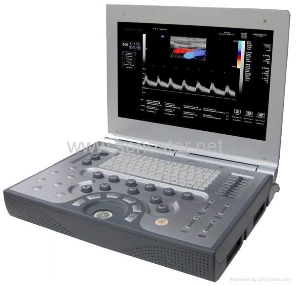
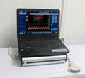
| Model: | SS-1200 |
|---|---|
| Brand: | - |
| Origin: | - |
| Category: | Electronics & Electricity / Electronic Instrument / Analysis Instrument |
| Label: | Doppler , Color Doppler , Ultrasonic |
| Price: |
-
|
| Min. Order: | - |
Product Description


USER INTERFACE | |
Ergonomic User Interface | |
• | Interactive soft-key controls, Track Ball |
• | Full keyboard supporting international character sets for text editing |
• | Optimized gain control with Eight time gain compensation ( |
• | Integrated unit with two transducer holders fixed on the console with locking design |
TECHNOLOGIES FOR PERFORMANCE | |
• | Frequency and Spatial Compound imaging |
• | Digital beam-forming from 12-bit A/D converters with TX/RX apodization for the best imaging resolution |
• | Multiple transmit focusing: 8 focal zones |
• | System dynamic range larger than 150 dB providing superb contrast resolution |
• | Digital image processing channels of over 1000 per image with synthetic aperture, spatial and frequency compounding. |
• | Optional Tissue Doppler Imaging |
• | Supports parallel receive beams to increase frame rate in color mode |
• | Optima parameter selection for Quick Scan and Optimized preset parameters for each application allow greater consistency and higher patient throughput |
• | Depth-dependent receive beam formation to increase penetration and image resolution |
• | Optimized sound speed adjustment to increase image resolution |
Programmable Technology Platform | |
• | Remote Services: Online customer services, maintenance and software upgrades available via the web |
• | Upgradeable platform: Optional upgrade to higher specification iMago portable models – P25 |
• | Networking: Modem, high-speed Ethernet, and Wireless 802.11b/g |
• | Tele-Radiology capable |
• | Vector Data Storage |
• | User programmable presets for specific clinical applications |
IMAGING MODES | |
B-Mode | |
• | Motion-Dependent Persistence with five user-selectable levels that reduces the noise and increases the contrast resolution |
• | Adaptive Contrast Enhancement with user-selectable brightness and contrast settings to increase the display sensitivity adaptively |
• | Panoramic imaging to see extended field of view of organs through a sweep of exam regions |
• | User-selectable contrast curves and contrast and color map setting to increase the display sensitivity interactively and for tissue contrast enhancement |
• | Adaptive Speckle Reduction to increase the contrast resolution and preserve the tissue structure |
• | Tissue Harmonic Imaging - THI Phase inversion for greater contrast and detail resolution, especially for patients considered technically difficult |
M-Mode | |
• | Specific user adjustable edge enhancement and dynamic range display |
• | User-selectable sweep speed for M-mode display with four settings |
Color Doppler (CFM) | |
• | 2-D flow processing to increase flow detection especially in highly noisy environments |
• | Intelligent tissue/flow detection with five user-selectable levels to reduce flow artifact and increase flow detection |
• | Flow persistence with five levels to increase flow signal to noise ratio |
Doppler Spectrum Mode (PWD) | |
• | Support spectral invert, 8 levels of baseline shift & wall filter selections |
• | Automatic baseline adjustment and waveform envelop detection |
Power Doppler Mode | |
• | Adjustable color map to increase color flow display sensitivity |
• | Support directional Power Doppler |
• | Mixed Mode Side-by-Side Live Display of combination B-Mode, B/Color Mode, B/Power Mode, B/Directional Power Mode |
Continuous Wave (CW) Doppler | |
• | Optional CW Doppler scan and spectrum |
IMAGE FEATURES | ||
Display | ||
• | Display Depth: up to | |
• | Display Gray Levels: 256; continuous variable contrast and frame rate of up to 300 frames/sec | |
• | Display dynamic range or compression: 45 to 90dB | |
• | Display Format: B, 2B, 4B, M, B/D, B/M, B/C, B/C/MC, B/D, B/P, B/C/D | |
• | Scanning Format: linear, parallelogram and trapezoid in linear arrays, and sector in phase and curved arrays | |
• | Side-by-Side image analysis to evaluate new features and parameter optimization, and Quadrant Imaging | |
• | Image Orientation: Left/right B mode reversal and up/down image invert; 90, 1800 rotation | |
• | Magnification: Zoom with pan capability in real-time or freeze B, C and M mode | |
• | Annotation: Allows the user to annotate anywhere on the image with pre-defined annotation list and anatomical body marks | |
• | Screen Display: Display of all patient and exam related imaging parameters, and on-screen documentation of image parameters in single / dual display modes | |
3D Ultrasound | ||
• | Freehand 3-D ultrasound |
|
• | Optional: 3-D display in B-mode and Color Doppler simultaneously |
|
Image Review | ||
• | CINE Review: Variable speed motion review and frame-by-frame review | |
• | Standard: 1,024 B-Mode frame and 170 seconds M-Mode data | |
• | Standard: 520 color frames and 380 seconds Doppler Spectrum | |
Image Management | ||
• | Standard: 160 GB hard disk drive for local image storing | |
• | ||
• | Standard: 2,000 Images w/ | |
• | Optional post processing of images for analysis | |
• | DICOM & PACs Compatible | |
MEASUREMENTS & CALCULATIONS |
| |||
• | Basic B-mode: distance, area and circumference, A/B ratio, angle, ellipse, volume, automatic IMT measurement for vascular applications |
| ||
• | Basic M-mode: velocity, heart rate, distance, slope, A/B ratio, time |
| ||
• | Basic Doppler: peak velocity/frequency, time, mean velocity, heart rate, RI, PI, S/D ratio, flow volume, area |
| ||
• | Basic Obstetrical Package: measurements include |
| ||
• | Basic Gynecology Package: measurements include: GYN and fertility protocol, size and volume of Ovaries, Uterus and Follicles, Cervix, Endometrium |
| ||
• | Basic Cardiac Package: measurements include |
| ||
• | Basic Urology Package: measurements include the volume of prostate, step wise and residual urine; reports cover: |
| ||
• | Knowledge based user friendly patient report editing and management: Obstetrical report, Gynecological report, Cardiac report, PV report, OB/GYN examination history |
| ||
CLINICAL APPLICATIONS |
| |||
Abdominal | Ob./Gyn. |
| ||
Breast | Small Parts |
| ||
Cardiac | Urology |
| ||
Musculoskeletal | Vascular |
| ||
TRANSDUCER OPTIONS | ||||
• | Linear array: 5.0 MHz, 7.5 MHz, 10 MHz (3.5 MHz to 14 MHz) | |||
• | Curved array: 3.5 MHz (2.5 MHz to 5 MHz) | |||
• | Trans-vaginal: 7.5 MHz (5 MHz to 10 MHz) | |||
• | Phased array: 2.5 MHz (2 MHz to 3.5 MHz) | |||
• | ||||
Member Information
| Sonostar Electronic Technologies Co., Limited | |
|---|---|
| Country/Region: | Guang Dong - China |
| Business Nature: | Manufacturer |
| Phone: | 18922710457 |
| Contact: | Chua (Market Manager) |
| Last Online: | 15 Jun, 2019 |
Related Products of this Company
-
UBox-9 Ultrasound B Scanner Box
-
SE-3B Digital Three Channel Color
-
Array Ultrasound Probe Scanner[coming
-
Sonostar hot sale cheap medical 3D 4D
-
UBook-8 Ultrasound B scanner(Notebook)
-
SS-6 Ultrasound B Scanner
-
Sonostar cheap trolley doppler
-
C200 Trolley Color Doppler Ultrasound
-
Sonostar new portable ultrasound box
-
Sector Ultrasound Probe Scanner[coming
